Why Does Milia Form and How Is It Removed?
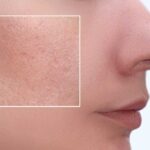
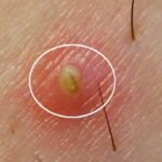
Milia are small, white or yellowish, firm and painless cysts, 1-2 millimetres in diameter, that form from the build-up of keratin beneath the uppermost layer of the skin. They are most often seen around the eyes, over the cheekbones, on the forehead and along the sides of the nose; in babies they are quite common during the newborn period. Unlike blackheads, milia are not a blocked pore; they are a mass of keratin trapped inside a closed sac. For this reason they do not come out when squeezed, do not melt away with creams and may not disappear on their own over time. So what exactly is milia, why does it form, how is it distinguished from a blackhead, can it be removed at home, which methods remove it in the clinic and what should be done to prevent recurrence? In this comprehensive guide, we address all these questions in detail.
What Is Milia?
Milia are medically defined as epidermal keratin cysts. In the epidermis, the uppermost layer of the skin, keratin, the main protein of dead cells, normally moves towards the surface and is shed. If this cycle is interrupted at any point, keratin becomes trapped inside a thin sac and hardens over time, forming a small, raised white spot. Because the cyst wall is completely closed, the content cannot escape; this explains why milia do not empty on their own. The lesions are painless, non-itchy and not inflamed. They feel firm and immobile when touched. At Zen Polyclinic milia are evaluated with magnified examination of the skin surface and distinguished from other lesions that look similar.
What Is the Difference Between Milia, Blackheads and Pimples?
These three lesions are frequently confused but their mechanisms differ. A blackhead (open comedone) is a blockage of the opening of the hair follicle with sebum and dead cells; because its surface is in contact with air it oxidises and darkens. A whitehead (closed comedone) is also a follicular blockage but its opening is closed and its content is mainly oil. Milia, on the other hand, are not a follicular blockage; they are a closed micro cyst filled with keratin and usually not connected to a follicle. Therefore the squeeze-and-empty methods used in acne treatment do not work for milia. In addition, milia do not become inflamed, do not turn red and leave no scar, whereas acne lesions can become inflamed and leave scars.
Why Does Milia Form?
The basic mechanism in milia formation is keratin not being able to reach the skin surface. The factors that trigger this are:
- Slowing of dead cell renewal (age, dryness, barrier impairment).
- Long-term use of thick, comedogenic creams and make-up products.
- Inadequate or excessively aggressive skin cleansing.
- Sun damage: a thickened upper layer blocks the flow of keratin.
- Trauma: burns, friction, the healing period after peeling, dermabrasion.
- Some medications (such as long-term topical corticosteroids) and genetic predisposition.
When these factors come together, the development of milia becomes easier, especially in areas with thin skin.
Distinguishing Primary and Secondary Milia
Milia are divided into two main groups. Primary milia develop spontaneously in healthy skin without any external cause; this is the type most often seen in newborns and around the eyes in adults. Secondary milia, on the other hand, appear after a situation in which the skin has been damaged: burns, blistering skin diseases, dermatological procedures, healing after aggressive laser or deep peeling, long-term use of steroid creams. Secondary milia are related to damaged sweat ducts and follicular structures trapping keratin during healing. This distinction affects the treatment plan: in secondary cases the underlying cause must be eliminated first, otherwise the lesions recur.
Why Does Milia Occur in Babies?
Milia are seen in about half of newborns in the first days of life; this is called milia neonatorum. Because babies’ sweat and oil glands are not yet fully mature, the flow of keratin is temporarily disrupted. The lesions most often appear as white spots on the nose, cheeks, chin and forehead. Milia in babies are completely harmless, cause no itching or discomfort and require no treatment. They usually disappear on their own within 2-6 weeks. The most appropriate approach during this period is to cleanse the skin gently with warm water, not to apply oily cream or baby oil and definitely not to try squeezing them. Medical assessment is recommended for lesions that persist for months or become widespread.
Why Is Milia Common in Adults?
In adults milia are usually a chronic process and less likely to disappear on their own. With age the rate of cell renewal slows, the skin barrier thins and the flow of keratin becomes more difficult. Added to this are thickening of the upper layer caused by sun damage, heavy moisturisers, eye creams, non-water-based sunscreens and make-up residues. Smoking and irregular skin care also accelerate the process. Adult milia may remain in the same place for years, so clinical intervention is needed. Knowing your skin type and choosing suitable products is a protective step. Our guide on how to identify your skin type offers guidance on this.
Why Is Milia So Common Around the Eyes?
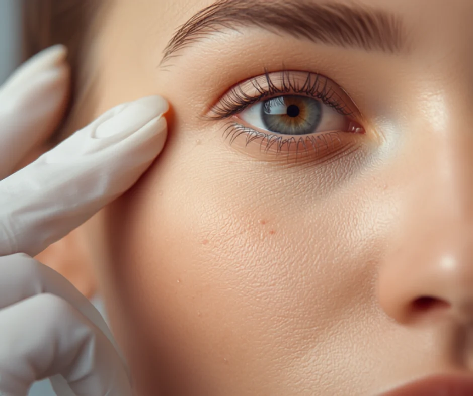
The eye area has the thinnest skin in the body and a low density of oil glands. When these two features combine, the flow of keratin is blocked more easily and the small cysts that form remain very close to the surface, so they become visible immediately. In addition, eye creams applied to this area are usually thicker in consistency and thin skin cannot carry this weight. Inadequate make-up removal in this area also contributes. White spots lined up on the lower eyelid and at the outer corner of the eye are the most typical appearance. Self-intervention in this area markedly increases the risk of scarring and discolouration because the skin is so thin.
What Are the Types of Milia?
Several subtypes have been defined clinically. Milia neonatorum is seen in newborns and is temporary. Primary milia appear in children and adults, especially around the eyes. Milia en plaque is a rarer picture characterised by milia clustered on a reddened base and is usually seen behind the ears and around the eyes. Traumatic (secondary) milia develop in healing areas after burns or procedures. In addition there are widespread forms of milia seen together with some genetic syndromes. Determining the correct subtype makes both the choice of treatment and the prediction of recurrence risk easier; therefore dermatological evaluation is important.
In Which Areas Is Milia Most Often Seen?
The most typical location is the upper half of the face: the eyelids and lower eye area, the cheekbone region, the forehead, the sides of the nose and the temples. Less frequently they may be seen on the cheeks, chin, ear and upper neck. Secondary milia depend on the area of damage; burn scars, graft areas, regions healing after laser or skin areas exposed to constant friction are examples. They have rarely been reported on the trunk, arms and genital area too. Location also provides information for differential diagnosis: for example yellowish plaques on the eyelid may suggest xanthelasma, and yellow-cream coloured bumps on the forehead and cheeks may suggest sebaceous hyperplasia.
Is Milia Contagious or Harmful?
Milia are not contagious; they do not pass from person to person and are not a bacterial infection. Nor are they a precancerous lesion; they are entirely benign. They do not cause pain, itching or inflammation. Therefore they do not require medically mandatory treatment; the request usually arises from aesthetic concern. However, in some situations medical advice is needed: rapid multiplication of lesions, clustering on a reddened base, accompanying itching or pain, size exceeding 3 millimetres or an uncertain diagnosis. In these situations differential diagnosis becomes important. If discomfort is only due to appearance, clinical removal is a safe and effective option.
Does Milia Go Away on Its Own?
In babies the answer is largely yes: milia neonatorum mostly disappear within a few weeks without treatment. In adults the situation is different. Primary milia can remain in the same place for months, sometimes years. The fact that the cyst wall is closed and the keratin has hardened makes natural elimination almost impossible. Some superficial lesions may open on their own after the upper layer thins with regular chemical peeling or retinoid use; however this should not be expected in every case. The general approach is this: wait and see in babies, while in adults clinical extraction with the right method is the fastest and safest solution.
Why Is Squeezing at Home Harmful?
Milia are covered by an intact layer of epidermis; therefore the content does not come out when squeezed. Forcing only damages the surrounding tissues. Attempts at home with needles, safety pins or sharp objects carry the risk of infection, permanent red marks, dark discolouration (post-inflammatory hyperpigmentation) and small scars. This risk is even higher around the eyes because the skin is very thin and healing marks remain noticeable. In addition, non-sterile instruments can lead to folliculitis or local infection. The correct approach is to open the epidermis over the lesion at a very small point with a sterile lancet and gently empty the content; this is a clinical procedure.
How Is Milia Diagnosed?
Diagnosis is mostly made by clinical examination. The physician evaluates the size, colour, distribution and consistency of the lesions and, if needed, examines the surface structure using a dermatoscope. Milia typically appear as 1-2 millimetre, firm, white-yellowish, dome-shaped lesions with no redness. History is also important: a newly started skin care product, a past burn, a laser or peeling procedure, long-term use of cortisone cream are all questioned. Rarely, in cases where the diagnosis remains uncertain or the lesions look atypical, a biopsy may be considered. Correct diagnosis is decisive in avoiding unnecessary treatments and managing expectations correctly.
How Is Milia Removed in the Clinic?
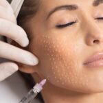
The most frequently used method that gives the fastest result is sterile extraction. The area is cleaned with an antiseptic and a topical anaesthetic cream is applied if needed. With a sterile lancet or a very fine needle, the thin epidermal layer over the lesion is opened by a fraction of a millimetre; then the keratin core is removed intact with a comedone extractor. The procedure takes seconds for each lesion; many lesions can be cleared in a single session. An antibiotic cream may be recommended afterwards. Healing is usually complete within a few days and leaves no scar. At Zen Polyclinic this procedure is carried out with minimal trauma and under sterile conditions so as to protect the skin barrier.
Milia Treatment With Plexr (Plasma)
In lesions that are numerous, very small or located in sensitive areas such as the eyelid, plasma technology is a strong option. Plexr provides very superficial and controlled vaporisation with plasma energy created by ionisation of air without touching the skin. In this way the epidermis over the milia is opened point by point or the cyst content is brought to the surface and eliminated. The advantage of the method is that heat spread to the surrounding tissue is very limited and it can be used safely in thin areas such as the eyelid. A thin crust forms after the procedure and flakes off within a few days. One session is usually sufficient; in widespread cases the session can be repeated.
Electrocauterisation and Laser Approach
In some cases, especially in deeply located lesions unsuitable for extraction, point opening with very low power electrocauterisation or fractional laser may be preferred. The aim is to open the tissue over the cyst with minimum damage and allow the keratin content to be eliminated. These methods require experience; applying too much energy can lead to hypopigmentation or the formation of small pits. Therefore power settings and pulse duration are determined carefully. Protective measures (eye shields) are used around the eyes. After treatment the area is kept moist for a few days, protected from the sun and crusts are not forcibly removed; this is the most important rule for avoiding scars.
The Role of Chemical Peeling and Retinoids
Chemical peeling and topical retinoids do not remove existing milia instantly; however, by accelerating the keratin cycle they reduce the formation of new lesions and bring existing ones closer to the surface. Salicylic acid is used for its oil-dissolving property, while glycolic and lactic acid are used for their effect of loosening the bonds between dead cells. Retinoids regulate epidermal cell renewal and provide long-term protection especially in recurrent milia. However, retinoids and acids should be used carefully around the eyes and with a physician’s advice; otherwise irritation and barrier impairment may develop. These treatments are usually planned as a maintenance approach after extraction and continued for weeks to months.
What Does Medical Skin Care Provide in Milia?
Professional care sessions are an important part of milia management. During medical skin care the skin is softened with steam or suitable solutions, the dead cell layer is reduced in a controlled manner and suitable lesions are emptied in a sterile way. In the same session the barrier is supported with a mask and moisturising appropriate to the skin type. Care carried out at regular intervals reduces keratin accumulation and thus lowers the frequency of recurrence. In addition, expert assessment can determine which home care products trigger milia. This is valuable information especially regarding eye creams and heavy sunscreens and allows the routine to be simplified.
Micro-Needling and Skin Renewal Treatments
In recurrent milia the long-term strategy is to increase the skin’s renewal capacity. Micro-needling methods such as dermapen stimulate collagen production by opening controlled micro channels and revitalise the epidermal cycle. Skin renewal treatments, on the other hand, ease the flow of keratin by reducing thickening in the upper layer. These methods are not direct milia treatments; they are supportive procedures that reduce the formation of new lesions. The treatment plan is made according to skin type and lesion density. An important note: because aggressive procedures can trigger secondary milia in some people, dose and depth settings must be determined under a physician’s supervision.
How Many Sessions Does Treatment Take and Does It Hurt?
With a limited number of lesions a single session is usually sufficient and the procedure takes 10-20 minutes. If there are widespread, numerous lesions it may be spread over 2-3 sessions so as not to stress the skin; 3-4 weeks are left between sessions. The pain level is low; patients usually describe a short-lived stinging or pinching sensation. Comfort can be increased by applying a topical anaesthetic cream 20-30 minutes before the procedure. Around the eyes the procedure is carried out more delicately and, if needed, several short sessions are planned. Slight redness after the procedure lasts a few hours; small crusts flake off within 3-5 days. It is possible to return to daily life the same day.
How Should Aftercare Be?
The healing period is short, but following the rules is important to avoid scarring. For the first 24 hours the area is not wetted or is cleansed very gently; afterwards a soft, soap-free cleanser is used. The antibiotic or healing cream recommended by the physician is applied regularly. Crusts must never be picked; they should be left to flake off on their own. Make-up should not be applied to the area until the crusts have gone. Sun protection is essential: SPF 30-50 protection prevents permanent discolouration in the healing area. Peeling, retinoids and scrubbing products are paused for a few days. Sauna, steam bath and swimming pools are not recommended for the first 3-5 days. If severe redness or discharge occurs, a physician should be consulted.
Does Milia Recur? How Is It Prevented?
An emptied lesion usually does not recur at the same point; however, in skin with a predisposition new ones may appear at different points. For this reason prevention is as important as treatment. The recommended approach: choosing light textured, non-comedogenic moisturisers and eye creams; always removing evening make-up; gentle chemical exfoliation 1-2 times a week; retinoid use with a physician’s advice; daily sun protection and regular professional care. Avoiding excessively rich care routines and unnecessary layers of products is also effective. Many of the protective principles that apply to blackhead and comedone removal are also valid for milia.
Product Choice: Which Ingredients Trigger Milia?
Heavy, occlusive (surface-sealing) ingredients increase the risk. Dense mineral oil, lanolin, solid fats and very rich eye creams can hinder the flow of keratin in thin-skinned areas. In particular, non-water-based, film-forming sunscreens may contribute to build-up around the eyes. Instead, forms labelled “non-comedogenic”, gels or light emulsions should be preferred. If silicone based primers and long-wear make-up products are used, the cleansing step should be in two stages (oil-based cleanser plus gentle facial cleanser). If new lesions are observed in the first 4-6 weeks after a product change, that product should be considered a possible trigger.
The Relationship Between Sun Protection and Milia
Chronic sun exposure thickens the stratum corneum, the uppermost layer of the epidermis, and damages elastic tissue. This thickened layer makes it harder for keratin to reach the surface and prepares the ground for milia formation. In addition, healing capacity decreases in sun-damaged skin and the risk of discolouration after procedures increases. Therefore daily sun protection is necessary both for prevention and for the period after treatment. Formula choice matters: light, mineral based or gel form products are more suitable around the eyes. Physical protection methods such as hats and sunglasses provide an additional advantage by reducing the product load.
The Role of Make-Up and Cleansing Habits
The most frequently neglected trigger of milia is inadequate make-up removal. Residues of eyeliner, mascara and concealer accumulate especially along the lower eyelid line. Rubbing this area firmly also worsens the situation by damaging the barrier. The correct approach is to apply eye make-up remover to a cotton pad, press it on the area for 10-15 seconds and then wipe gently. Rather than using waterproof products every day, using them only when needed is recommended. In addition, washing make-up brushes regularly is a detail often overlooked in recurrent lesions. A simple and consistent routine is more protective than a complex and heavy one.
Conditions That Mimic Milia
Small white-yellowish bumps are not always milia. Syringoma are skin-coloured papules arising from sweat ducts, symmetrically located on the lower eyelid. Xanthelasma appears as yellowish, soft plaques on the eyelid and may be related to lipid metabolism. Sebaceous hyperplasia consists of yellow-cream coloured bumps with a depressed centre. A closed comedone, on the other hand, is a follicular blockage and its content comes out when squeezed. Their treatments differ; a wrong diagnosis leads to unnecessary procedures and disappointment. Therefore medical evaluation is definitely recommended, especially in numerous, symmetrical or atypical lesions.
When Should You See a Physician?
Evaluation should not be delayed in the following situations: rapid multiplication of lesions, increase in size, accompanying redness or itching, discharge, crusting or bleeding, lesions clustering on a reddened base and uncertainty about the diagnosis. In addition, professional support is needed for lesions that have previously been self-treated and left marks. Milia in newborns that persist for months or become widespread should also be evaluated. Seeing a physician is valuable not only for treatment but also for correct diagnosis and identification of triggers. At Zen Polyclinic the evaluation also includes skin type analysis and review of the home care routine.
Realistic Expectations: What Can and Cannot Be Done?
Clinical removal performed with the right method usually clears existing milia lesions in a single session and without scarring. This is a fast and satisfying result. However, there are also things that cannot be done: no method guarantees that new lesions will not form; the tendency is individual and partly genetic. Creams do not dissolve existing cysts. Interventions at home increase the risk of scarring. In addition, in recurrent cases maintenance treatment (exfoliation, retinoids, regular professional care) must be continued; a one-off intervention does not mean a permanent solution. Knowing these limits is the most important element in increasing treatment satisfaction and eliminates unrealistic expectations.
Milia Treatment at Zen Polyclinic
At Zen Polyclinic the approach to milia starts with a detailed skin examination and differential diagnosis; the type and location of lesions and possible triggers are evaluated. In suitable cases removal is performed in a single session with sterile extraction; in sensitive areas such as the eyelid and in widespread lesions plasma technology (Plexr) may be preferred. After treatment, the risk of recurrence is reduced with a care plan that supports the skin barrier, suitable product recommendations and, if needed, a retinoid or exfoliation protocol. We serve with our expert team in our clinics in Istanbul Ataşehir. You can create an appointment for a personalised assessment.
Conclusion
Milia are benign, non-contagious small cysts that form when keratin cannot reach the skin surface and becomes trapped inside a closed sac. While they usually disappear without treatment in babies, in adults they can be persistent and require clinical intervention. Trying to remove them at home by squeezing or with a needle carries the risk of scarring and infection; the correct solution is sterile extraction or, in suitable cases, plasma based methods. To reduce the risk of recurrence, light textured products, regular gentle exfoliation, sun protection and professional care are important. If you are not sure about the type of lesions or if they are multiplying rapidly, we recommend having an individual assessment with a medical examination.
Frequently Asked Questions (FAQ)
What is milia?
Milia are small, white or yellowish, firm and painless cysts 1-2 millimetres in diameter that form from the build-up of keratin beneath the uppermost layer of the skin. Because they are a keratin mass trapped in a closed sac, they do not come out when squeezed.
What is the difference between milia and blackheads?
A blackhead is a blockage of the hair follicle with sebum and dead cells and darkens because it is open and oxidises. Milia are not a follicular blockage but a closed micro cyst filled with keratin; they do not become inflamed and do not resolve with acne treatments.
Why does milia form?
Slowed renewal of dead cells, use of thick and comedogenic creams, inadequate cleansing, thickening of the upper layer due to sun damage, trauma such as burns or peeling, and genetic predisposition all make milia formation easier.
Does milia go away on its own?
Milia neonatorum seen in babies usually disappears within 2 to 6 weeks without treatment. In adults the lesions may remain for months or years; because the cyst wall is closed natural elimination is very difficult and clinical intervention is needed.
Can milia be removed at home by squeezing?
No. Because the lesion is covered by intact epidermis, the content does not come out when squeezed. Using a needle or sharp object at home carries the risk of infection, permanent red marks, dark discolouration and scarring; the risk is especially high around the eyes.
How is milia removed in the clinic?
The most common method is sterile extraction: the thin layer over the lesion is opened by a fraction of a millimetre with a sterile lancet and the keratin core is removed. In numerous lesions or sensitive areas such as the eyelid, plasma methods like Plexr may be preferred.
Does milia treatment hurt and how many sessions does it take?
The pain level is low; a short stinging sensation is described and a topical anaesthetic cream increases comfort. A single session is sufficient with a limited number of lesions; in widespread cases 2 to 3 sessions may be planned 3 to 4 weeks apart.
Does milia recur and how is it prevented?
An emptied lesion usually does not recur at the same point, but new lesions may appear in predisposed skin. Light textured and non-comedogenic products, regular gentle exfoliation, retinoids with a physician’s advice, daily sun protection and professional care reduce the risk.